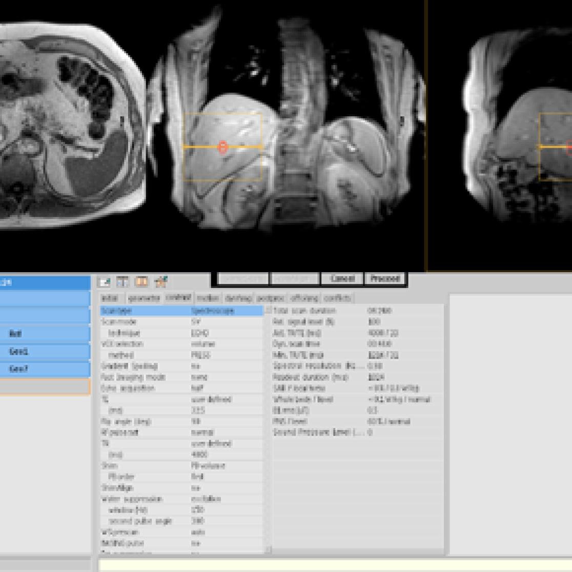
Imaging – non-invasive investigation of metabolism
Developing non-invasive imaging of liver inflammation
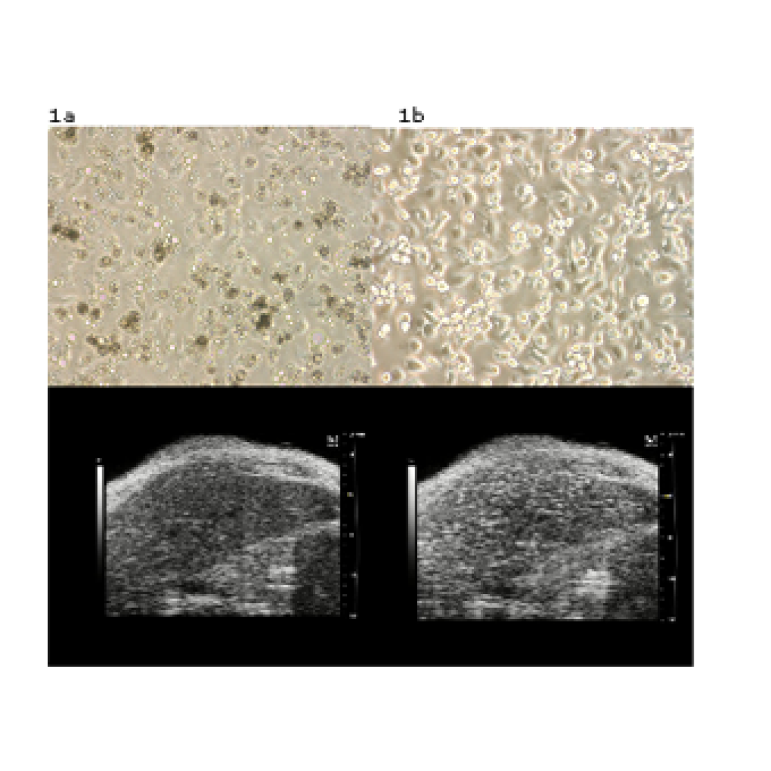
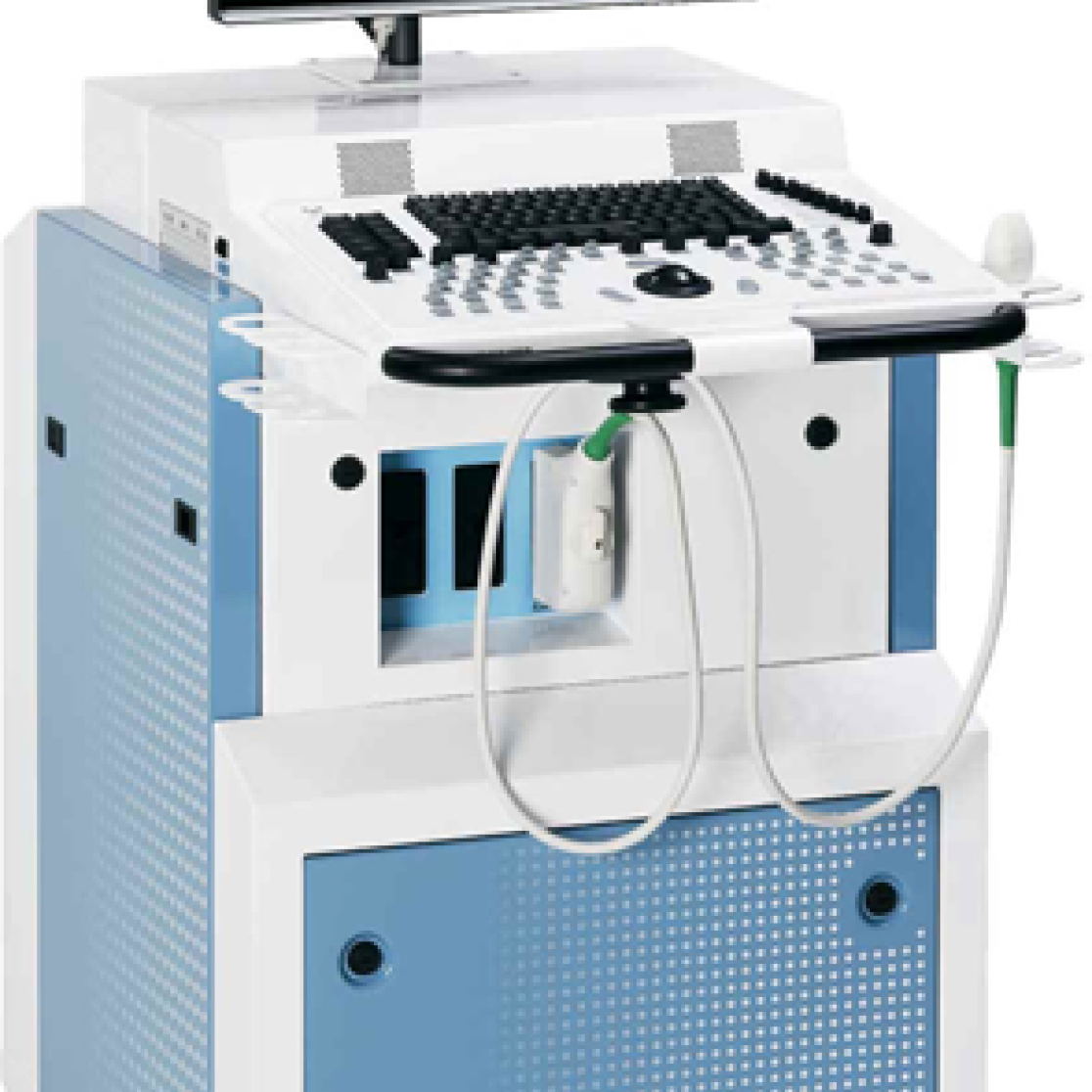
At present, liver biopsy is the gold standard for diagnosing liver inflammation but its use has several limitations including sampling error, patient discomfort and a risk of serious complications. We are currently investigating the possibility of using Ultra-Sound Imaging echogenic loaded Bone Marrow Derived Macrophages, as a non-invasive imaging technique.
Information: Dr. Ronit Shiri-Sverdlov, Department of Molecular Genetics
Figure: Microscope view of cultured bone marrow derived Macrophages and an ultrasound view of a mouse liver a) unloaded macrophages b) macrophages loaded with a ultragenic particles c) ultrasound of a mouse liver after injecting unloaded macrophages d)
Modulating the hematopoietic lineage by Bone Marrow Transplantation
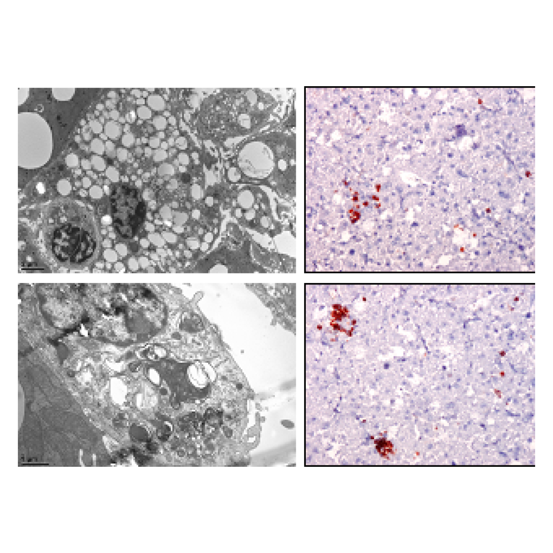
To determine the specific contribution of the hematopoietic cells, to diseases progression, we transfer Bone Marrow from mice with genetic modulation of the relevant pathway into recipient mice. Analysis of the liver is done by Electron Microscopy and Immuno-Histochemistry.
Figure: Electron Microscopy and Light Microscopy photos from a mouse liver. a) +c) EM photos of a Kupffer cell loaded with fat b)+d) Light microscopy photos of the liver stained for a Kupffer cell marker.
Imaging bone structure by High Resolution peripheral Quantitative Computed Tomography (HR-pQCT)
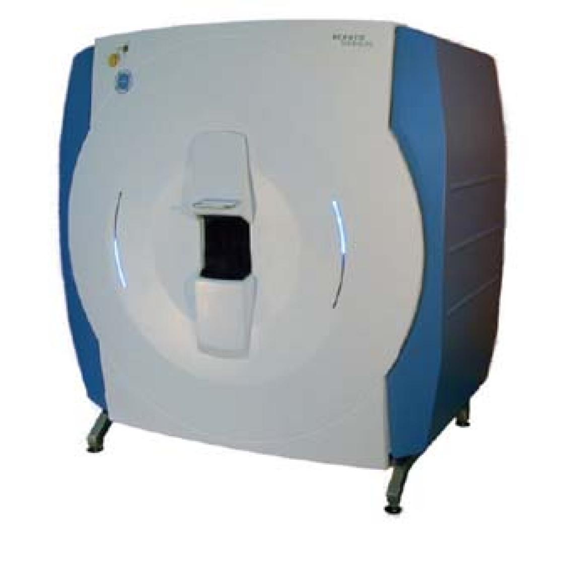
High resolution peripheral quantitative computed tomography (HR-pQCT) scanning allows in-vivo assessment of 3D bone density and architecture on the micro-scale at a resolution of 82 micrometer in the distal radius and tibia. Based on these assessments, bone strength can be calculated with FEA (finite element analysis). Currently, the only HR-pQCT scanner (XtremeCT by Scanco) in the Benelux is located at the department of De Maastricht Study and this state-of-the-art technique is now being used in several research projects.
Information: Prof. Dr. Joop van den Bergh, Department of Movement Sciences and Internal Medicine (Rheumatology).
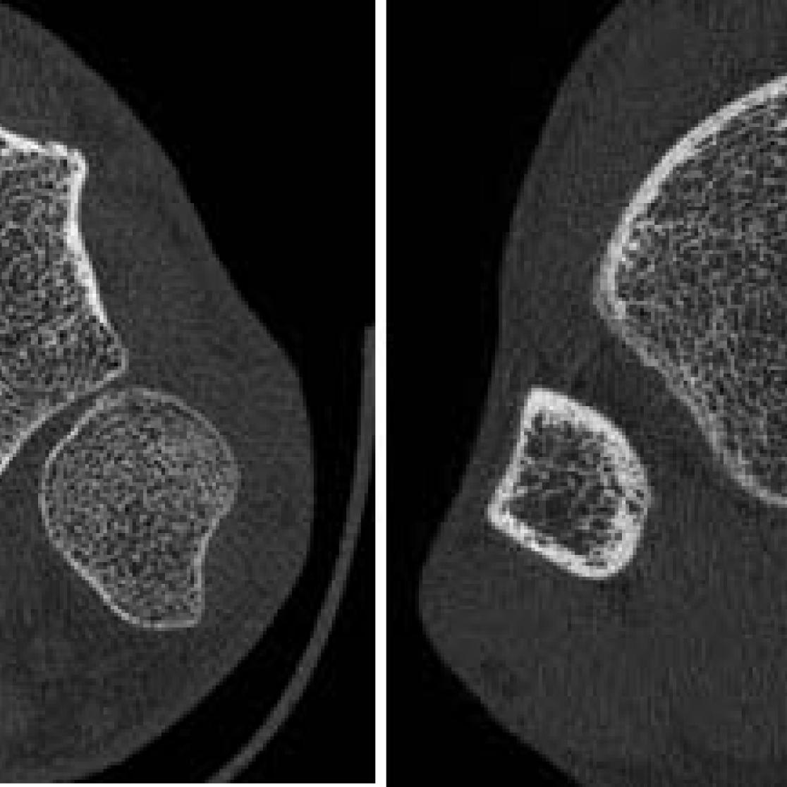
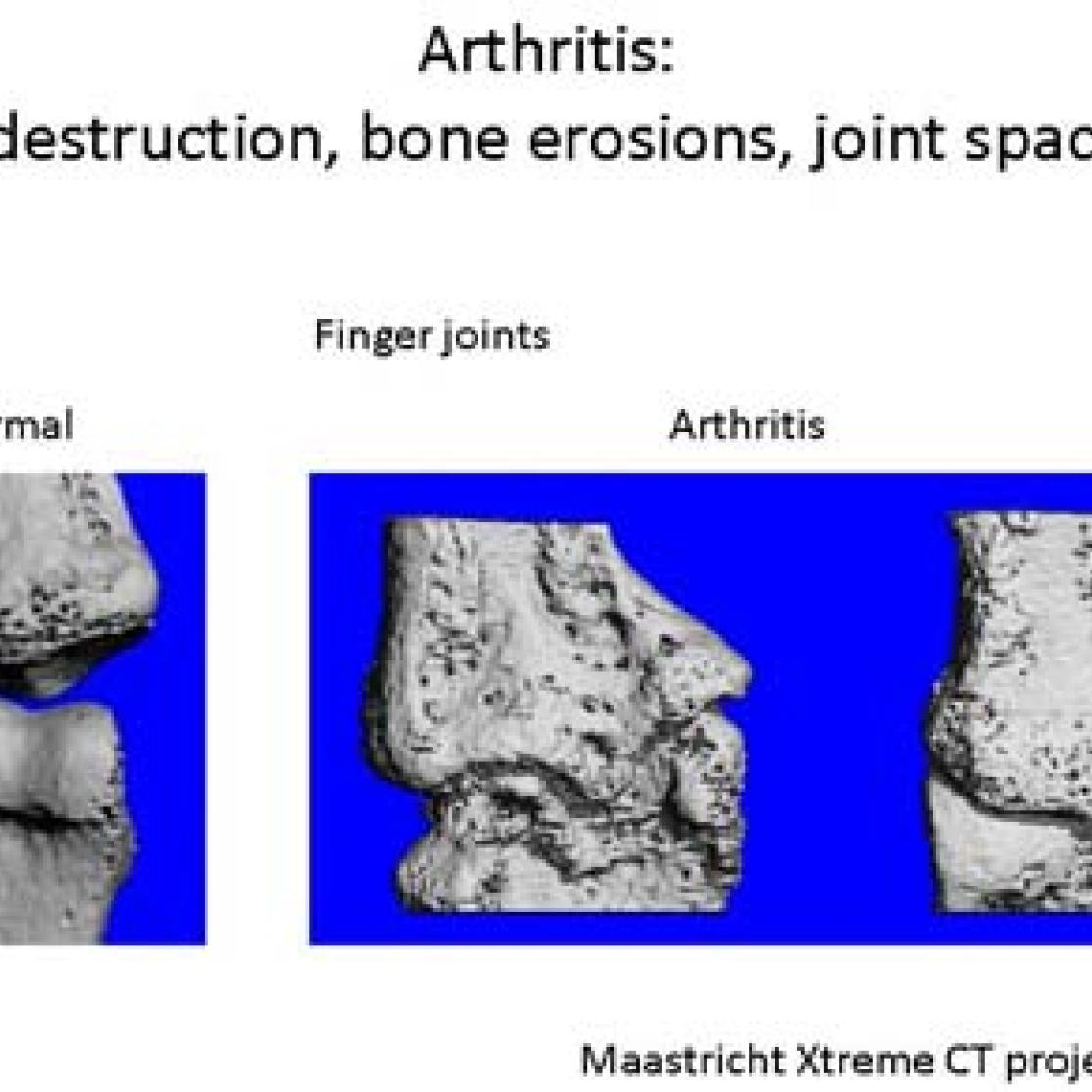
Bone erosions in the finger joints of patients with rheumatoid arthritis and deformations in osteoarthritis of the finger joints can be assessed using the HR-pQCT in combination with advanced image analysis techniques.
Figure: 3D reconstruction of a normal and osteo arthritis finger joint scanned by HRpQCT
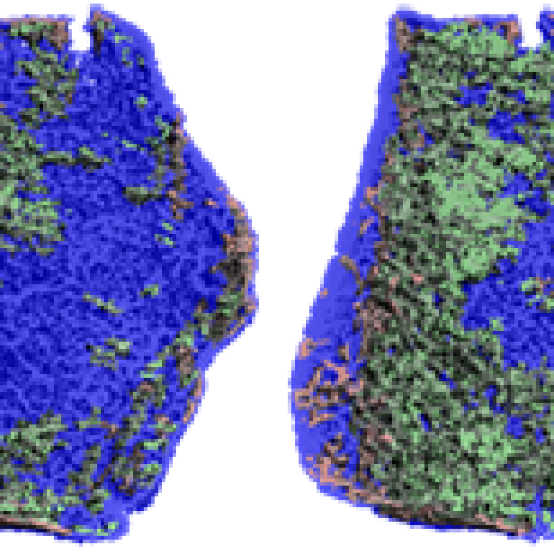
The HR-pQCT scanner is also used to study the fracture healing process of distal radius fractures. Regions of bone gain and bone loss can be visualized separately by subtraction
Figure: Visualization of the locations of bone gain (green) and loss (red) during the fracture healing process
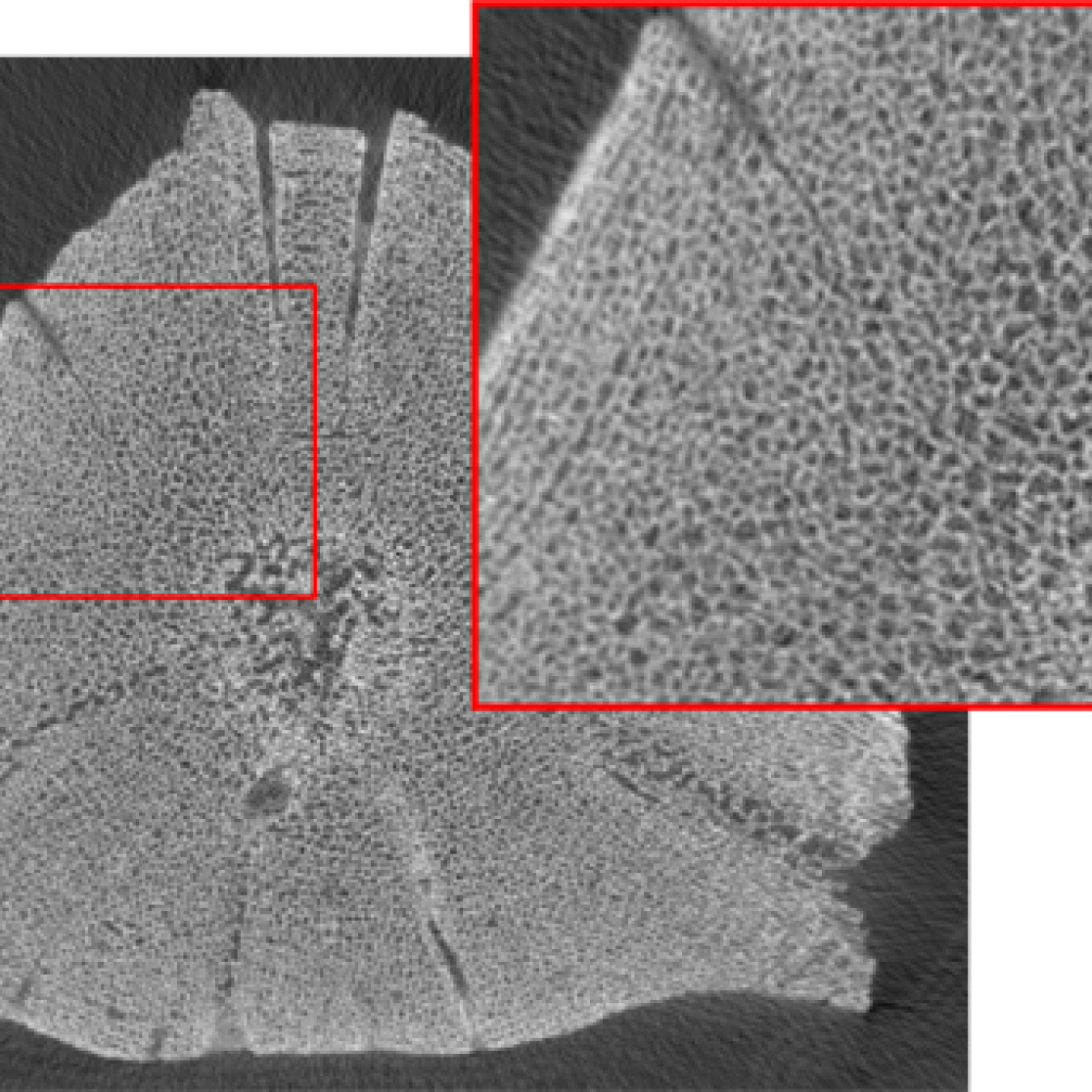
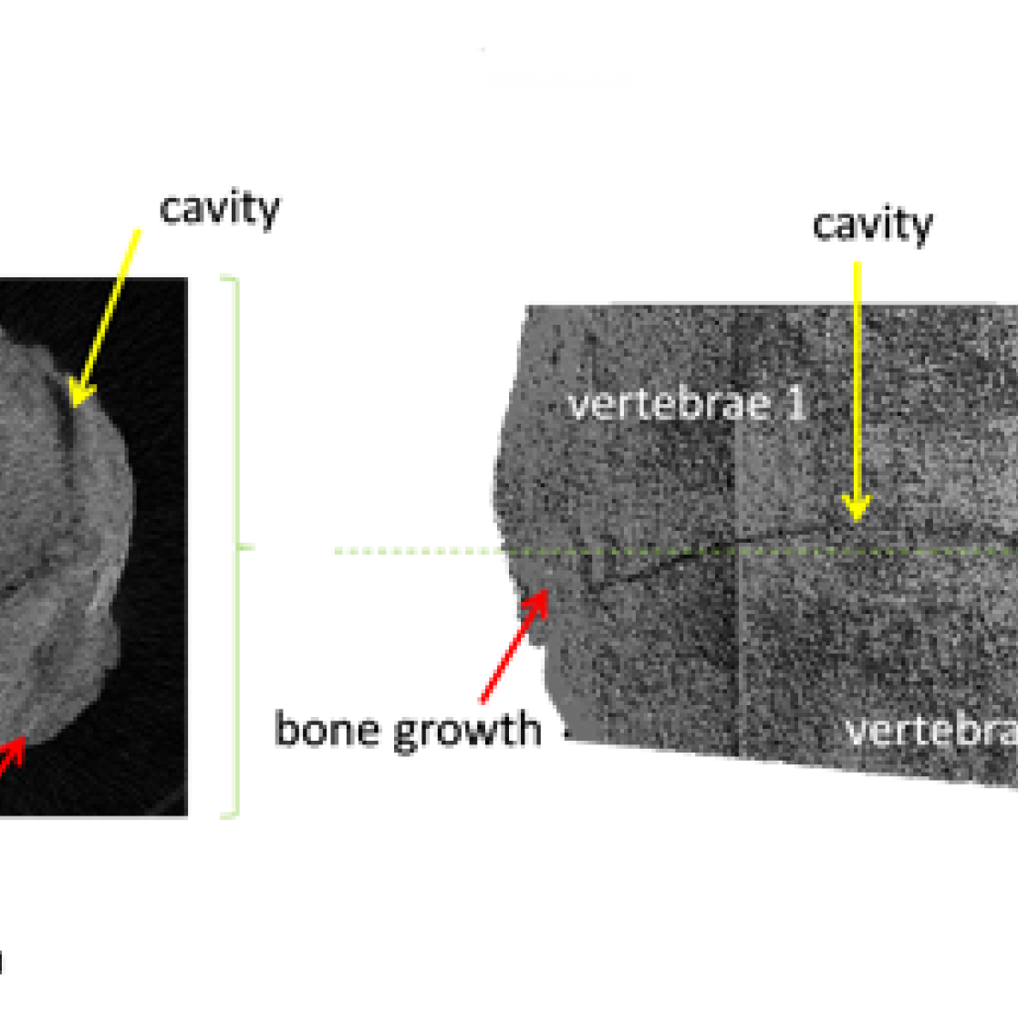
Occasionally, the HR-pQCT is also used for special requests, e.g. in the CSI Mosasaurus project - a collaboration with Natuurhistorisch Museum Maastricht. In this project, the bone structure of the famous 66 year old fossil Bèr (found in the southern part of Limburg) was assessed and two connected vertebrae were scanned by HR-pQCT in order to determine intervertebral bone growth.
Muscle function and “In-Vivo” anatomy
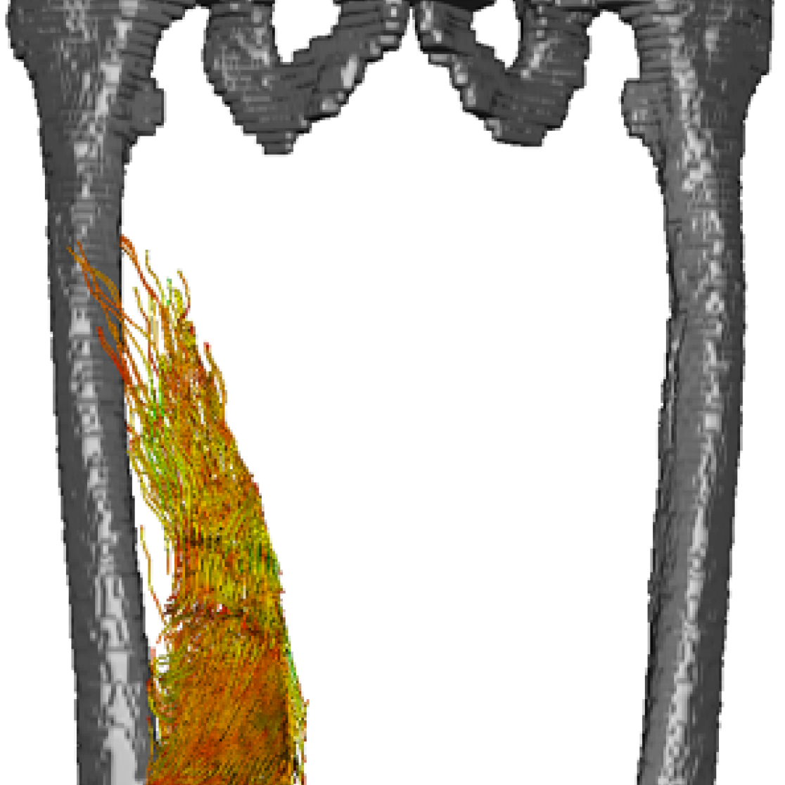
Muscle capacity is determined from dynamometry experiments. The HPL has a BIODEX III dynamometer which is used to characterize the force and power capacities of muscle groups. Muscle anatomy is a major determinant of muscle capacity. DTI-MR Imaging is used to conduct “In-Vivo” anatomy and capture the volume, fiber length, physiological cross sectional area (PCSA) and bony attachment sites of muscles. This information is vital to the translation of the dynamometry data into the fundamental length-force and force-velocity characteristics of muscle.
Information: Dr. Kenneth Meijer, Department of Movement Sciences
Non-invasive investigation of metabolism
Non-invasive investigation of metabolism is possible by Magnetic Resonance Spectroscopy (MRS). For example, ectopic fat content can be determined in skeletal muscle, the heart or liver.
Information: Dr. Vera Schrauwen-Hinderling, Departments of Radiology and Human Biology
by 1H-MRS. Similarly, high-energy metabolites, such as ATP or Phosphocreatine can be quantified by 31P-MRS in these organs.
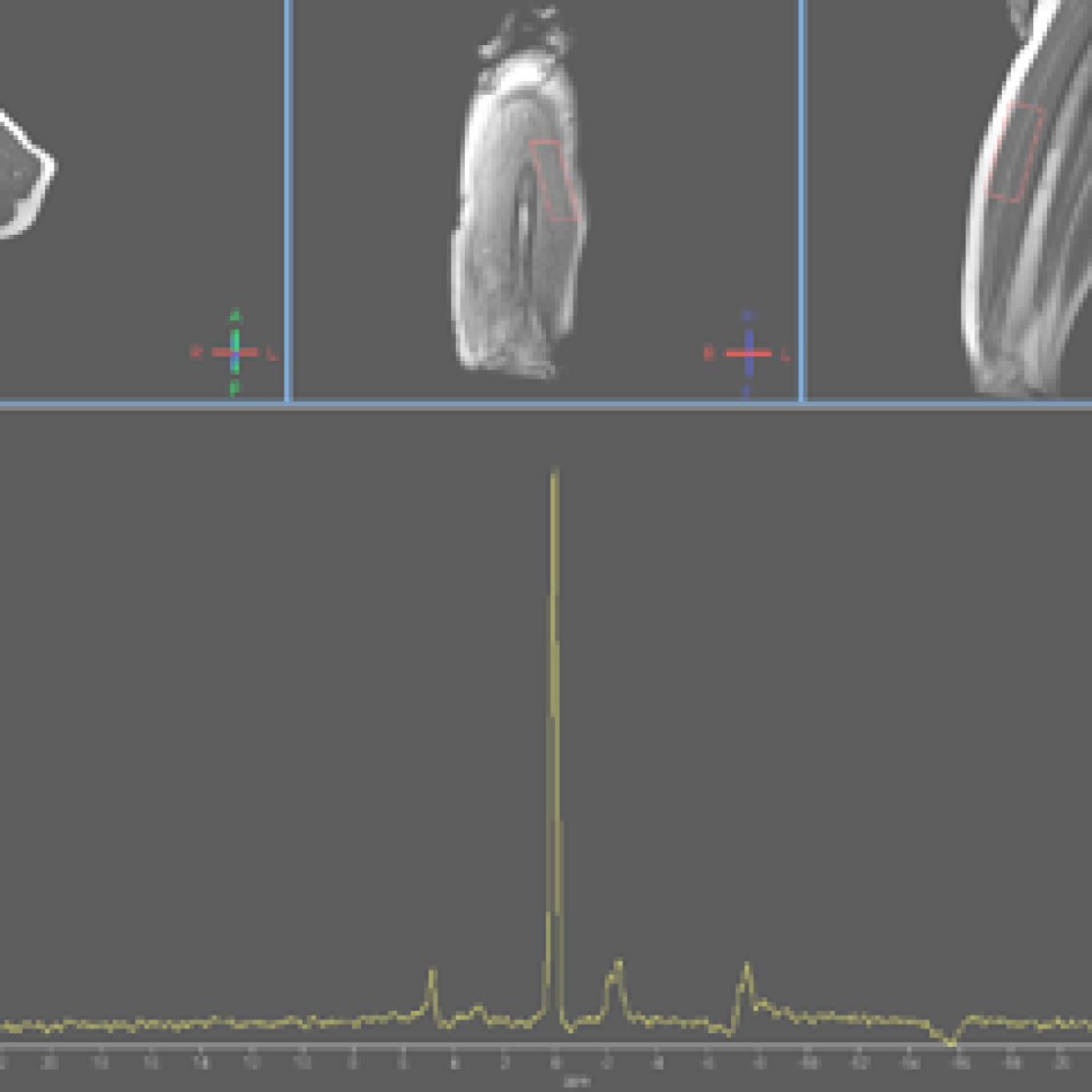
These techniques are valuable tools in evaluating the effects of physiological interventions and are currently applied in the evaluation of a physical activity programme and several dietary interventions. Currently, 13C-MRS is set-up, which is opening new possibilities in the characterization of lipids.
