Stroke
Crossroad
Research theme: Neuroimaging
Clinical pillar: Stroke
We apply various advanced vascular magnetic resonance imaging (MRI) techniques and image analysis methods to patients with stroke, vascular cognitive impairment and its interaction with brain tissue and function, to answer fundamental and clinically relevant neurological questions, and thus obtain a better understanding of the underlying mechanisms of stroke. Activities are in collaboration with the School for Cardiovascular Diseases CARIM.
Unique contributions and highlights
Cerebrovascular MRI in cerebral small vessel disease (cSVD)
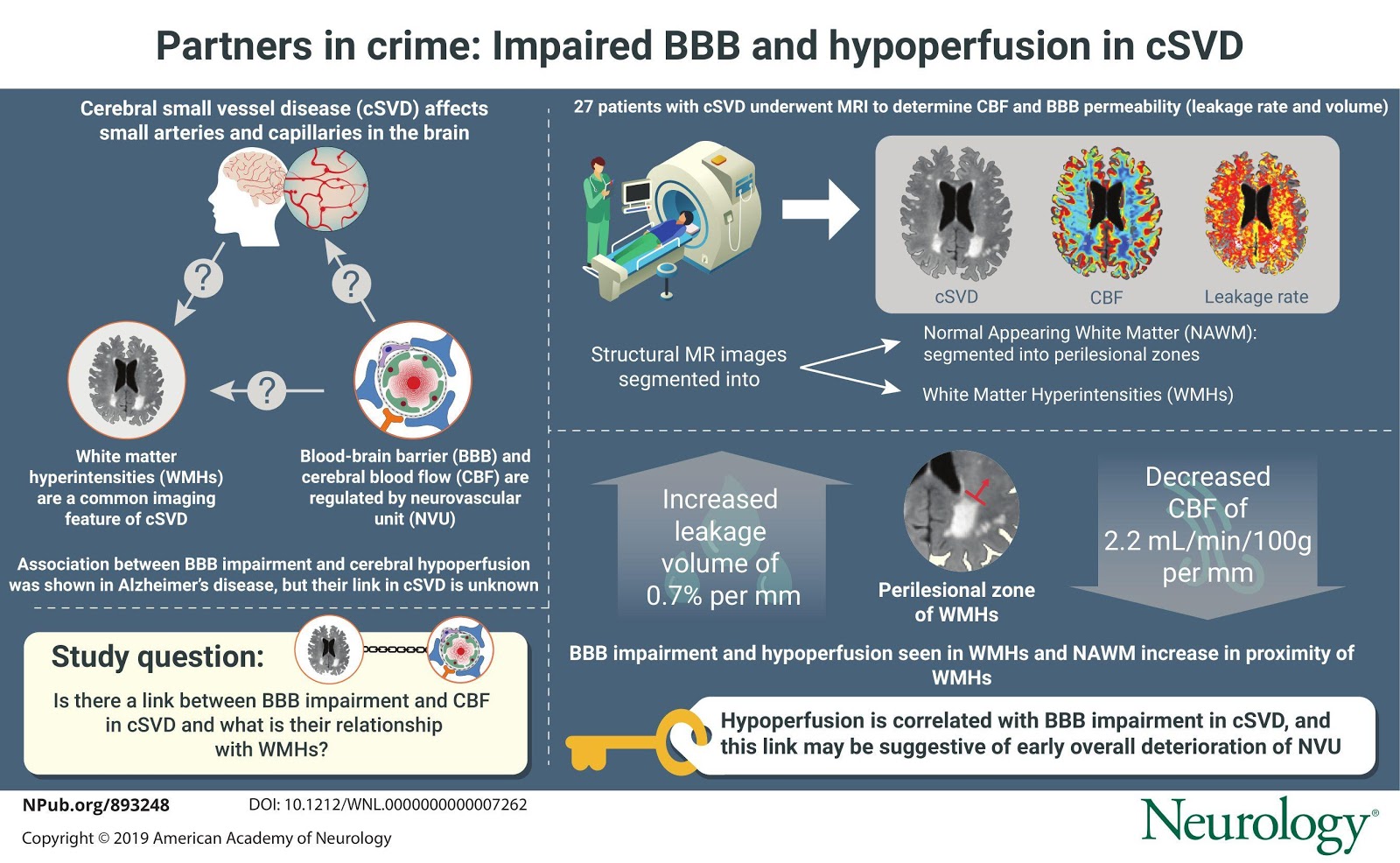
cSVD is a common microvascular pathology underlying burdensome diseases like lacunar stroke, and is an important contributor to cognitive impairment and dementia. The exact pathophysiology of cognitive deterioration in SVD remains unclear.
We developed several advanced cerebrovascular MRI techniques, and demonstrated in patients with cSVD 1) subtle blood brain barrier (BBB) leakage (Zhang et al., Neurology. 2017;88(5):426-432), 2) altered microvascular perfusion and parenchymal diffusivity (Wong et al., Neuroimage Clin. 2017;14:216-221) and 3) an impaired neurovascular unit (Wong et al., Neurology. 2019;92(15):e1669-e1677).
In 2019, a H2020 grant was awarded to an international consortium, in which Maastricht will apply cerebrovascular MRI techniques in vascular dementia and heart failure. Furthermore, a translational project, in which the relationship between BBB permeability and post-stroke epilepsy will be investigated, was funded in 2019 by ZonMW and the National Epilepsy Foundation.
Ultra-High field MRI
Ultra-high field MRI (≥7T) holds promise for cerebrovascular pathology, as it allows for better visualisation of brain structure and function. We recently demonstrated that the pulsatile blood flow through the lenticostriatal arteries can be precisely measured using 7T MRI and reveal effects of arterial stiffness due to aging. (Schnerr et al. Front Physiol. 2017;8:961)

