Division of Nanoscopy Facilities
The division of Nanoscopy is equipped with cutting-edge scientific equipment.

Division of Nanoscopy Infrastructure
Cryo-electron microscopy is the only way to study cellular processes close to the in vivo situation. In order to do so, we have implemented a full workflow, starting with life cell imaging of cells or tissues followed by rapid cryo-fixation to preserve the structure. Cryo-light and Cryo-electron microscopy will resolve the 3D structure at nm resolution by means of electron tomography.
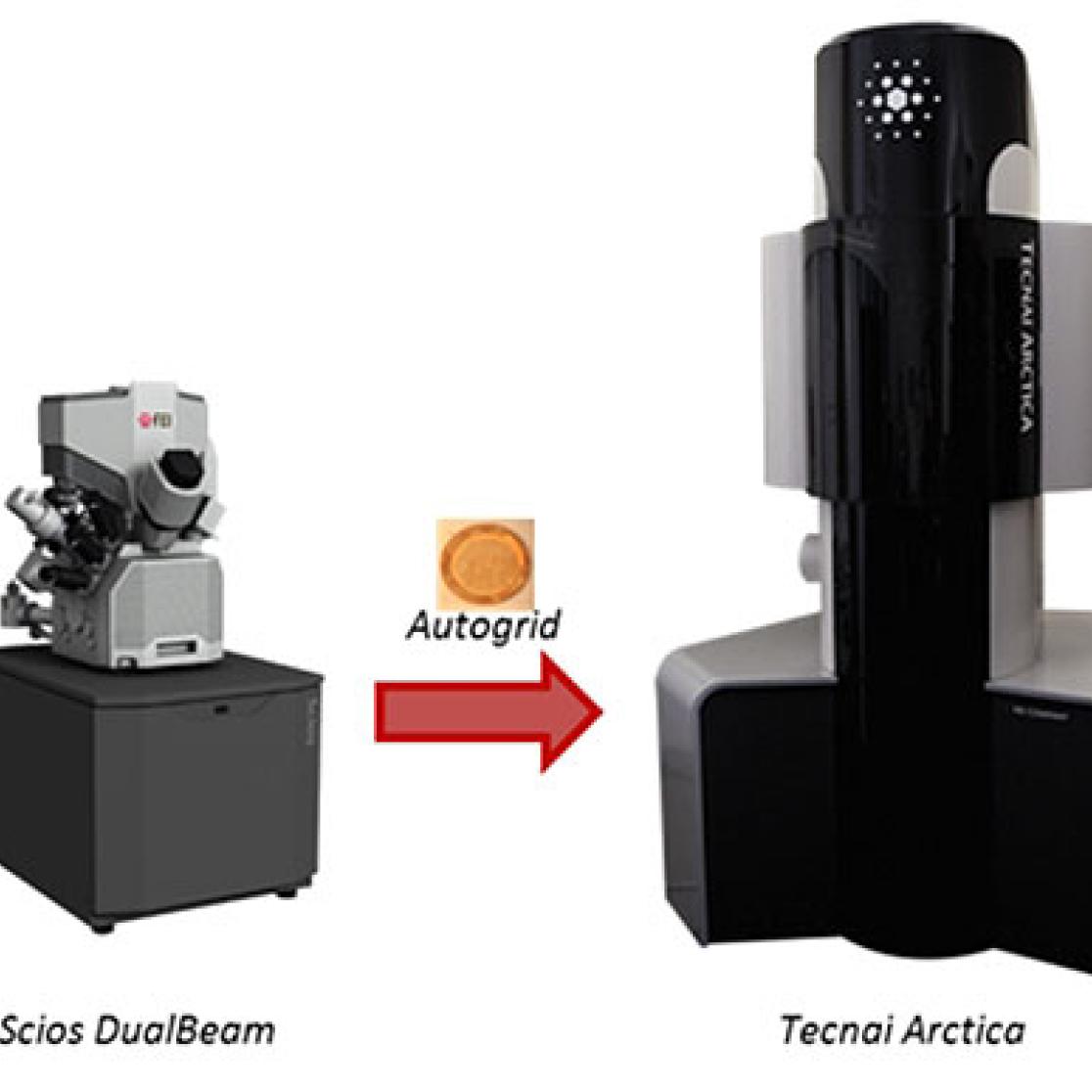
Our full cryo-correlative workflow for ‘Selected volume tomography’
Step 1
The workflow starts on the CorrSight with live cell imaging of cell cultures on an EM carrier, which can be used throughout the complete workflow in order to prevent loss of orientation and sample contamination or destruction. Once a specific event or location has been identified by means of light microscopy and preserved by means of cryo-fixation, the vitrified sample can be re-examined on the CorrSight in a dedicated cryo-stage in order to precisely determine the position of interest.
Step 2
After transferring the sample to the Scios DualBeam, the identified region is thinned down to the appropriate thickness of about 200-300 nm without artifacts using the focused ion beam. The correlative MAPS software allows the transfer of all data from the CorrSight such as positions of interest and recorded images to the Scios DualBeam in order to know exactly where to prepare the lamella for subsequent high resolution cryo-TEM imaging.
Step 3
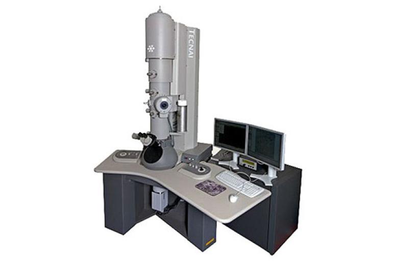
After thinning the samples to the proper thickness, the grids are loaded in the autoloader on the Tecnai Arctica for high resolution cryo-tomography. The Tecnai Arctica is equipped with FEI’s high sensitive Falcon direct electron detector for low-dose imaging, FEI tomography software for automated low-dose tomography and EPU software for automated single particle data acquisition.
More equipment
In addition to the CorrSight, Scios DualBeam, Tecnai Arctica, the Division of Nanoscopy also has the following equipment, which can be seen in the slideshow:
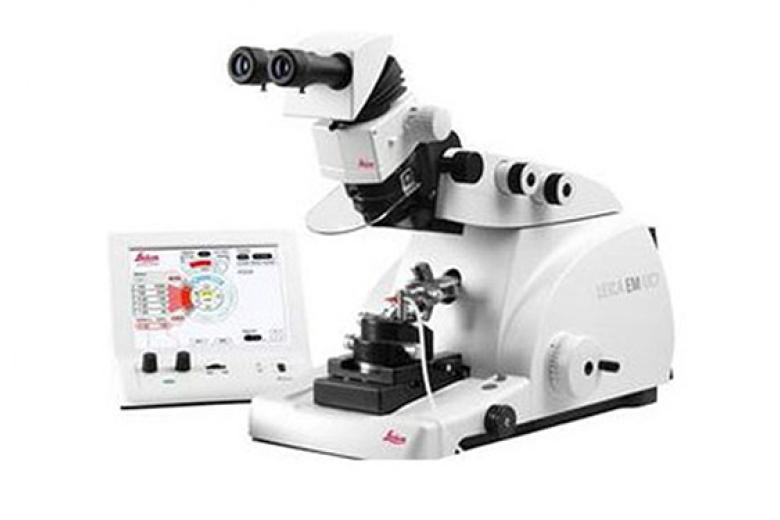
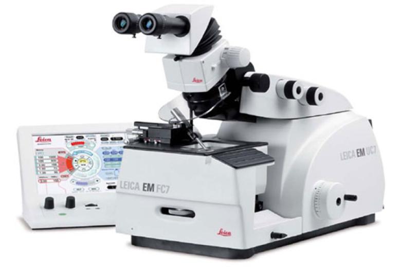
- Ultramicrotome Leica EM UC7
- Cryochamber Leica EM FC7
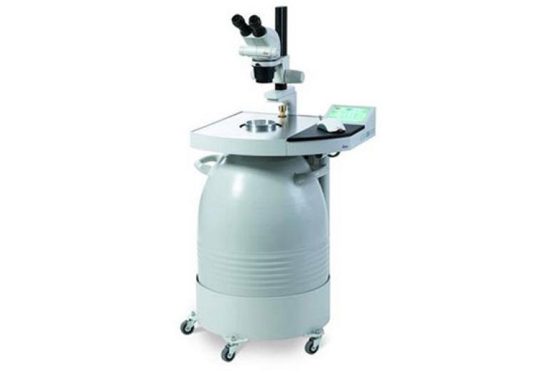
- Leica EM AFS2
- Combined iCorr fluorescence light microscope module and the FEI Tecnai electron microscope
M4I office wing
The M4I office wing has been designed with the same open and transparent look and feel as our labs. Based on C.O.R.E. collaborative open research education. C.O.R.E. requires a transparent and open environment for both laboratories and offices. M4I has invested heavily in an innovative and open environment for collaborative research. Research and office space is shared by scientist from very different backgrounds and disciplines, ranging from the fundamental sciences, technology and engineering as well as clinicians. In line with the CORE philosophy of Maastricht University the infrastructure is primed for researchers to cross the boundaries of their own disciplines and stimulate each other to excel in translational imaging science.
