Mapping disease and health at cell level with new microscopic technique
Scientists at Maastricht University developed a technique to better map different cell types and their location in tissues. This innovation allows scientists to better understand disease processes such as arteriosclerosis and cancer. The first findings with this new technique were published in the leading scientific journal Cell Metabolism.
Larger palette
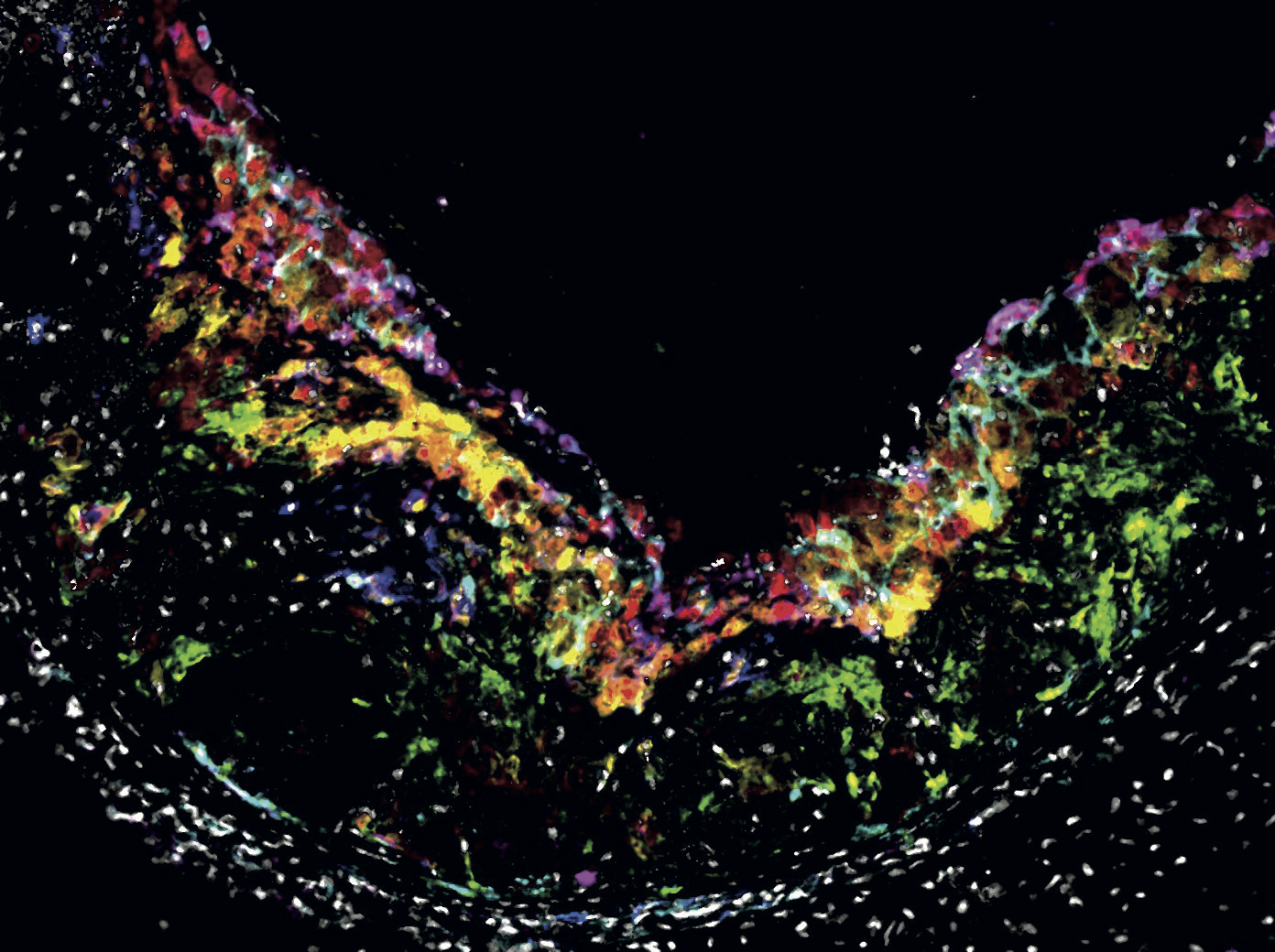
Many disease processes involve many different types of cells. In order to properly understand how diseases develop, it is important to know which cell types tissues are made up of. Until recently, researchers could distinguish a maximum of five cell types at a time using microscopic imaging, by giving cells each a different color. UM scientists developed a new method that can image up to fifteen different cell types. They used a technique called multispectral microscopy. With this type of microscopy, they look at cells like at white light passing through a prism showing all the colors of the rainbow: the cells are not visible as a single color, but as a unique combination of different colors. This way they can observe more cells at the same time, and better understand of which cell types certain tissues are made up off.

Communicating
The Maastricht scientists' method can determine the location of each cell type in the tissue ánd shows which cell types are located in each other's vicinity (known as cell communities). Neighboring cell types are known to influence each other: for example, inflammatory cells in tumor tissue counteract the defense of tumor cells, with major negative consequences for the course of the disease. "We can now investigate how certain cell types communicate with each other and whether they are involved in the disease process," explains UM researcher Pieter Goossens (Experimental Vascular Pathology). In addition, the researchers coupled the multispectral microscopy technique with the mass spectrometry imaging (MSI) of the M4I Institute. "This new method now allows researchers to map the molecular environment of the cells and its influence on their behavior, and that provides leads for medical treatment."
Also read
-

-
Better chances for cancer in the liver
Our liver is a special organ: if you cut away part of it, in most cases a new piece of liver will grow back. If someone has cancer in the liver, the affected part of the liver can be surgically removed. But you can only do this if at least 30% of the liver remains. For many patients whose remaining...

-
Diagnosis of skin cancer via quick scan a step closer
Determining whether a suspected spot on the skin is a basal cell carcinoma - the most common form of skin cancer - can be done in a large number of cases with a scan of the skin instead of an invasive biopsy. This has less impact on the patient, is faster and can lead to cost savings in healthcare.
