Helping surgeons and pathologists with the iKnife
When removing a tumour, it’s not always easy for a surgeon to decide whether all of the cancer cells have been taken out. Obviously, removing too much healthy tissue is not desirable either and sparing organ tissue is of paramount importance. The ‘intelligent knife’, or iKnife, can play an important role in this in the future. Researchers from the Maastricht M4I institute, led by Prof. Ron Heeren, are key partners in an international consortium that will validate the method, and have started the second phase of the experiments (in vivo) in a Dutch hospital last fall.
First molecular operating room of the Netherlands in Maastricht
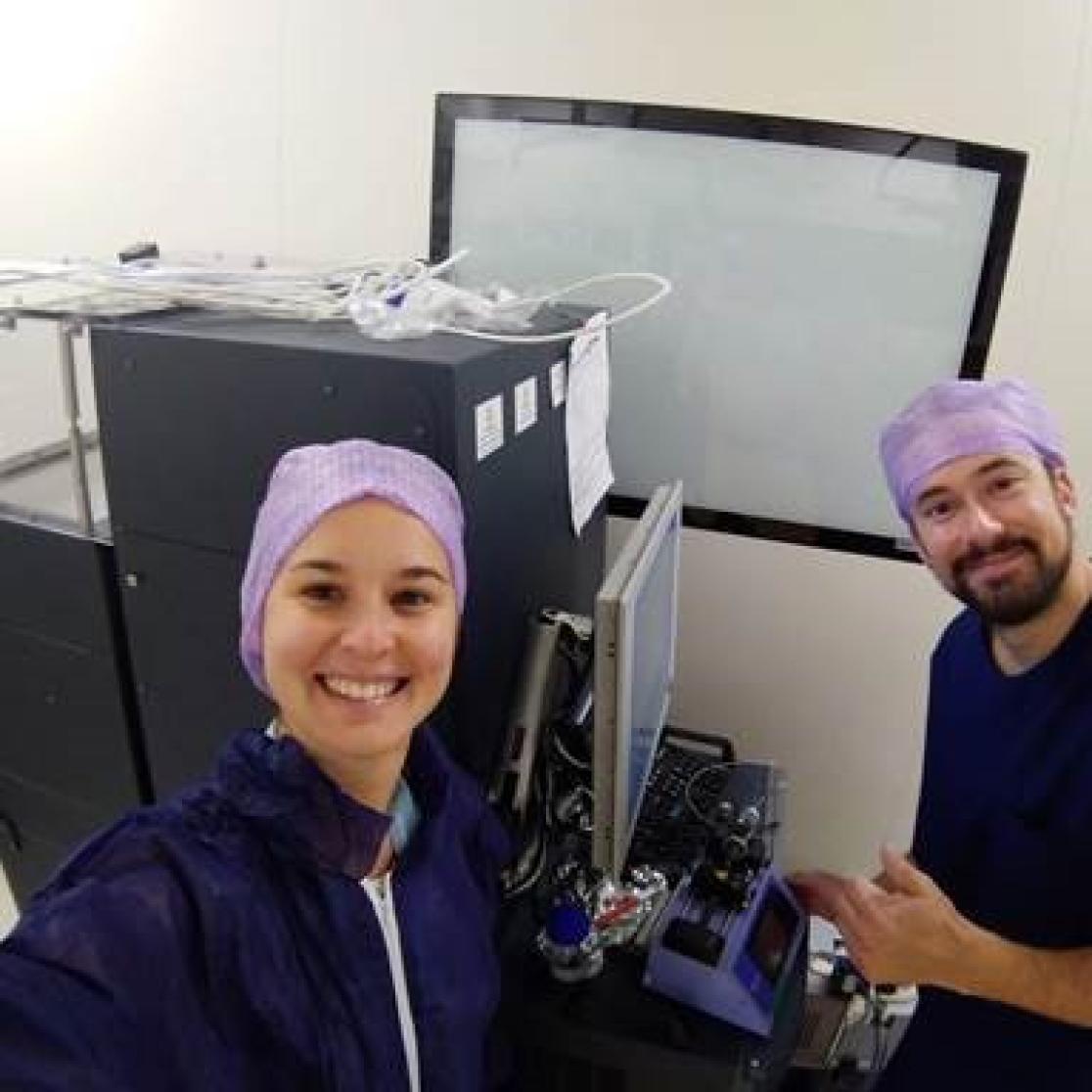
Dr. Tiffany Porta is an assistant professor at M4I and an expert in mass spectrometry imaging (MSI). She heads the iKnife project, which is all about ultimately taking the technique to the clinic. Close collaboration with the Maastricht University Medical Centre+ (MUMC+) is key, since it is there that the first ‘molecular operating room’ of the Netherlands is implemented. The surgeons, pathologists and clinical team involved are very enthusiastic. Prof. Steven Olde Damink, surgeon at MUMC+, was one of the early adopters of the technology here in Maastricht. Tiffany Porta and Pierre-Maxence Vaysse, the first PhD student working on the project, have been working hand and glove with the clinical team to establish the entire workflow, from patient selection to tissue analysis, and have been collecting patient tissue since January 2017. So far, about 300 patients have contributed tissue samples to the research. This is enabling the tissue databases to be built.
How the iKnife works
“The knife is heated and creates smoke when it makes contact with the tissue during the surgery. This smoke contains molecules and it’s like a fingerprint of the tissue. The ‘molecular profile’ is very rich in information, including tissue-specific profiles that discriminate between the tumour and surrounding tissue – data that’s not available to the naked eye of the surgeon”, Porta explains. In the end, the technique would not replace anyone’s role, she stresses, but it would help in making a more accurate diagnosis. “This electrosurgery would simply make operations faster and make sure that all cancer tissue is removed during the initial operation.”
International multi-centre study to prove the robustness of the method
Before being accepted worldwide, the technique first needs to be validated and, for that, a multi-centre study has been initiated by an international consortium. The other academic partners are Queen’s University in Kingston, Canada and Imperial College London, UK. Waters is the company that holds the patent for the iKnife and is the private partner in the consortium working on improving the technology. Currently, a database with tissue from breast cancer patients is being built up in the three academic centres. “If we can use the Maastricht database to classify tissue in London and Kingston, the method is robust”, Porta explains.
Implementation of in vivo measurements in Maastricht
All the current activities with the iKnife have the sole purpose of research. The next step is to go to the operating room to collect and compare the molecular profiles in vivo during surgery. The implementation of the system in the operating rooms has started with head and neck surgeries in December 2018 by Porta’s research group in close collaboration with the clinical teams involved in the different projects. “First, we have to prove that it works in vivo and then it will be easier to push forward”, Porta concludes. The validation of the database is the crucial step towards ultimately having a clinical trial take place.

Also read
-
Green school playgrounds boost concentration and wellbeing
Children at schools with green playgrounds are better able to concentrate and display more social behaviour. This is the conclusion of a follow-up study within the long-running project The Healthy Primary School of the Future .
-
Reconnecting the brain and body: how to control devices with your thoughts
Can you control a robotic arm with your thoughts? Paul Weger (MHeNs) studies this to give back independence to patients with neurological conditions.
-
Ron Heeren appointed fellow of the Netherlands Academy of Engineering
Professor Ron Heeren, distinguished university professor at Maastricht University (UM) and director of the Maastricht MultiModal Molecular Imaging Institute (M4i), was appointed as a fellow of the Netherlands Academy of Engineering (NAE) on Thursday 11 December.