How the coronavirus infects our small intestines
These are exceptional times, also in science. Whereas publishing an article in the top journal Science normally takes at least a year, a group of researchers from Utrecht, Rotterdam and Maastricht managed to do it in six weeks. Seven employees from Prof. Peter Peters’ lab worked on it full-time, sometimes late into the night. The group was able to depict, with the help of advanced microscopy, how the coronavirus invades the cells of the small intestine, after which it multiplies rapidly. It is the scientific substantiation of gastrointestinal symptoms caused by the virus and underlines the importance of good hand hygiene after visiting the toilet or handling incontinence products, among other things. Peter Peters discusses the research, the virus and the vaccine.
The expertise of professor of nanobiology Peter Peters lies in unravelling cell structures down to the smallest level. With the aid of ultra-strong microscopes, he normally studies the tubercle bacillus, for example, which is responsible for one and a half million deaths each year from tuberculosis. Just like the coronavirus, the bacterium has ‘spikes’ that stick out. “These complex spikes in the tubercle bacillus consist of about fifteen different proteins.” Actually, you should look at the end of a spike like a key. The virus can only penetrate a human cell if a lock is found into which the key can fit. Lung cells, for example, have that lock. In scientific terms, it’s called the ACE-2 receptor. The group with which Peters collaborated for this research showed that the virus is capable of attaching to this receptor, which is also present in intestinal cells. “In eight hours, the virus creates hundreds of new viruses that can newly infect neighbouring intestinal cells and the intestinal cell itself dies off.” This was worthy of publication in the top journal Science.
Mini organs play the leading role
‘Mini organs’, also called ‘organoids’, were used for the research. In a laboratory, an organoid that has the properties of a real organ can now be grown from a stem cell. Medicines, for example, can be tested on a piece of intestine, without a human having to suffer. Or you can bring the coronavirus into contact with a mini intestinal organ and see what happens. The lab of Prof. Hans Clevers at the Hubrecht Institute in Utrecht took care of the organoids for this study, among other things; the lab of Dr Bart Haagmans at the Erasmus University Medical Center in Rotterdam brought them into contact with the coronavirus; and then the preparations of the organoids fixed in strong water came to Maastricht, where a high-tech electron microscope is located. “Fixing them in strong water makes the virus inactive and allows us to work with it safely”, says Peters.
An unprecedentedly fast process
In his lab in Maastricht, the electron microscope was running day and night, operated by Kevin Knoops. On his laptop in his Maastricht apartment, the professor was able to study the 100,000-times magnified images of how the virus penetrates the intestinal cells. Colleague Raimond Ravelli developed a way to ‘sew’ all the images of the preparations together, creating a large map of an organoid, similar to Google Maps. And while the data was still coming in, the Science paper was put in the pipeline. “I've never experienced that before”, Peters looks back. The speed of the reviewers was also unprecedented, and within a few days a second version was made, which was accepted for publication. The gold medal in science was won. But more importantly, the group showed how sensitive our intestinal cells are to the coronavirus.
Why washing hands with soap is essential
This leads to the hypothesis that the strong immune reaction that is triggered in the intestines when the virus appears may be so intense that the entire immune system ‘goes berserk’. “If you see such an abnormal immune reaction in the lungs, a flood of white blood cells enters the lungs, which doesn’t leave enough room to exchange gases and thus absorb oxygen.” These and more follow-up research questions need to be addressed, and are probably already being addressed here and there around the world, partly as a result of the Science article. What the research already provides now, in addition to the explanation for the gastrointestinal symptoms of many COVID-19 patients, is more awareness of the contagiousness of stools. “No intact virus particles have been found in stools yet, but the genetic material in the virus has been found. It is almost certain that the stools of COVID-19 patients are still contagious. So, this requires a lot of caution after every visit to the toilet, as well as among staff in nursing homes who change incontinence products. Good hand hygiene is essential.” Because the virus has a layer of fat that holds it together, washing with soap is very important, he stresses. “Soap destroys fat, causing the virus to fall apart and become harmless.” Finally, the study also provides a ‘proof of principle’; it shows how hundreds of thousands of new substances and all current medicines can be tested with mini-organs, in the search for a treatment for COVID-19.

Artist and scientist David S. Goodsell made this reality-like image of the corona virus (CSB Protein Data Bank; doi: 10.2210/rcsb_pdb/goodsell-gallery-019)
The key to the vaccine
And then there is the vaccine, which the whole world is yearning for so that ‘normal life’ can be resumed, older people can be hugged again, and more. To arrive at a vaccine, it is essential that you know exactly how the virus works. Chinese researchers have already published 3D animations of the structure of the ‘spikes’ of the coronavirus. “These show which proteins are on the ends of the spikes, but also that part of each spike is coated with sugar molecules. That’s what the virus does to protect itself from the human immune system. Antibodies, for example from a vaccine, cannot attach to proteins with sugar structures. So, with your vaccine, you want to access elements of the spike that are not coated with sugar molecules; the human body will create antibodies to attack this, which in fact disables the key of the coronavirus. Then it won’t be able to penetrate human cells.”
Or a medicine cocktail instead?
That doesn’t sound so simple and Peters can wake us up from the dream—it isn’t. Although hundreds of groups of scientists across the globe are working on it around the clock, he thinks the chances are very slim that there will be a vaccine that protects us against the coronavirus in a year's time. “We’ve been looking for a vaccine to protect against AIDS for 30 years, a vaccine for tuberculosis and malaria for 100 years.” The professor thinks that a cocktail of different medicines will be found sooner for the treatment of COVID-19. “Just like with the AIDS virus, where for years the symptoms have been suppressed very successfully with a combination of medicines. That cocktail intervenes in three stages of the virus. Even if a mutation occurs in one stage, you still have two other drugs that will work. According to Darwin's principles, it is impossible for all three to mutate at the same time. That’s why it works so well.” And speaking of mutating—it is expected that the coronavirus will also mutate in the future and that a new variant of the vaccine will therefore be needed every year, just as with the flu shot, in order to provide people with the best protection possible.
Another UM contribution
In order to examine viruses (or parts of them) under electron microscopes, they need to be frozen in a special way in liquid ethane. The university scale-up CryoSol-World at the Brightlands Maastricht Health Campus, which was co-founded by Peters, is developing the Vitrojet, which is the successor to the Vitrobot. This device, developed by Peter Frederik and his colleagues, is currently being used worldwide in coronavirus research. “It has appeared in a number of good publications”, says Peters proudly. “It’s great to see that UM can also contribute in this way to research into the coronavirus and the neutralising antibodies that can inhibit the virus”, says Peter Peters.
Also read
-
Evidence-based health tips for students: supermarket psychology
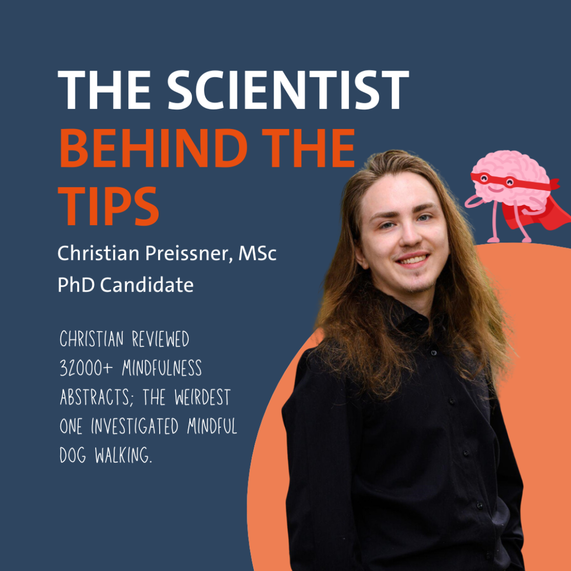
In the upcoming months, we’ll share tips on Instagram for our students on how to live a healthier life. Not just a random collection, but tips based on actual research happening at our faculty. The brains behind this idea are L ieve Vonken and Gido Metz, PhD candidates at CAPHRI, the Care and Public...Students,Students going the extra mile andUM news
-
Evidence-based health tips for students: the science of love and sex
In the upcoming months, we’ll share tips on Instagram for our students on how to live a healthier life. Not just a random collection, but tips based on actual research happening at our faculty. The brains behind this idea are L ieve Vonken and Gido Metz, PhD candidates at CAPHRI, the Care and Public...Students,Students going the extra mile andUM news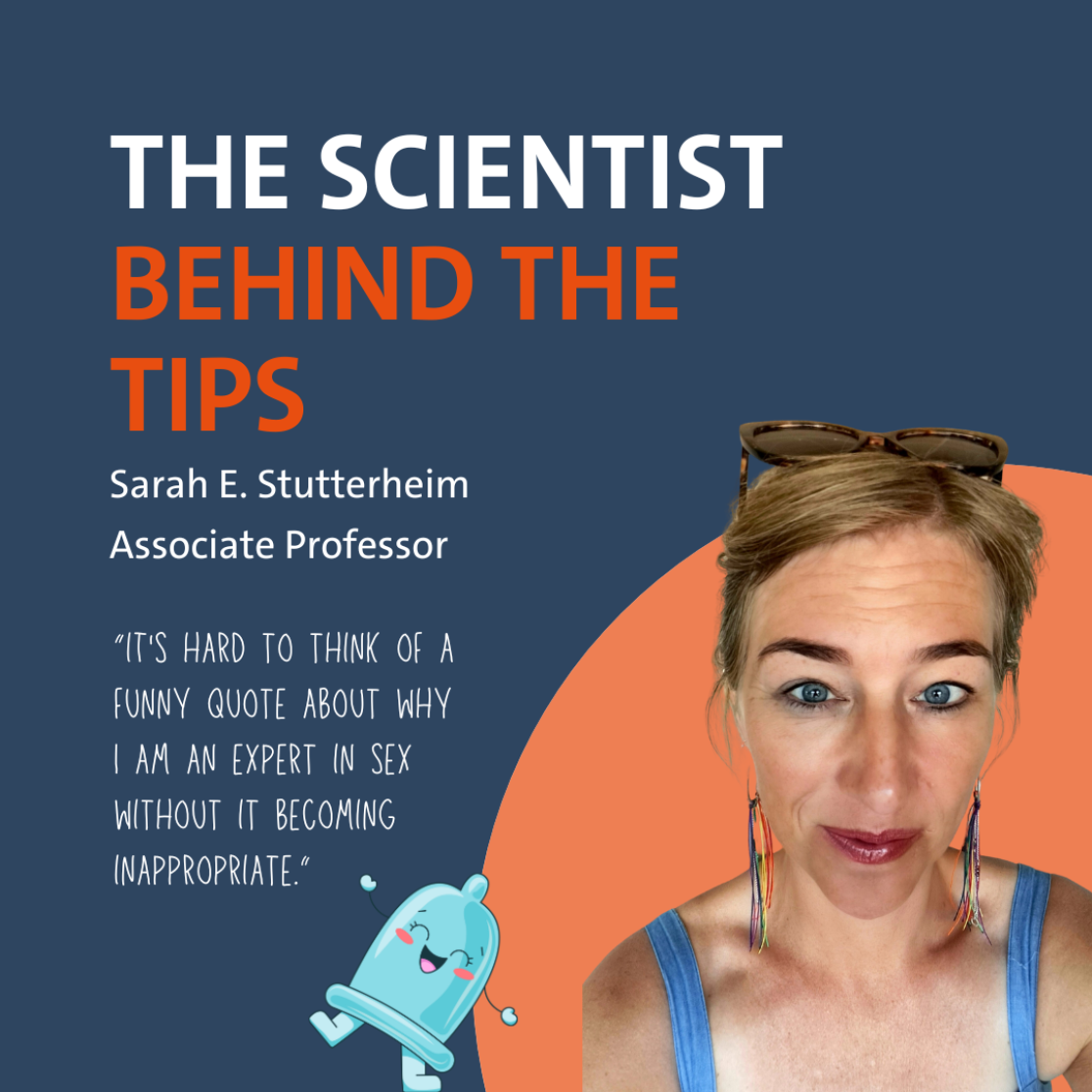
-
Evidence-based health tips for students: the science of mindfulness
In the upcoming months, we’ll share tips on Instagram for our students on how to live a healthier life. Not just a random collection, but tips based on actual research happening at our faculty. The brains behind this idea are L ieve Vonken and Gido Metz, PhD candidates at CAPHRI, the Care and Public...Students andStudents going the extra mile