Seeing things you cannot see

Berta Cillero Pastor is an Associate Professor and group leader at the MERLN Institute for Technology-Inspired Regenerative Medicine. Her research is centred around mass spectrometry (imaging)1 to gain insights into molecules in cells and tissues for biomedical research. With this technique, changes at the molecular level become visible that would otherwise be imperceptible2. One of her ongoing studies focuses on the early detection of osteoarthritis3, a research endeavour of notable significance given the prevalent occurrence and substantial healthcare costs linked to osteoarthritis.

Nice to talk to you! What is your role at MERLN?

Berta Cillero Pastor: "I am group leader at the MERLN Institute. I completed my postdoctoral work in Amsterdam at the AMOLF Institute. In 2015, I joined Maastricht University to establish the mass spectrometry imaging CORE lab platform at the M4i Institute, a ground-breaking concept for Maastricht. Over time, as my focus shifted more and more towards my independent research line, I decided to pursue opportunities in that direction at MERLN. In my current position, with a double affiliation with M4i, I have the privilege to have access to the world-leading infrastructures of the M4i and MERLN institutes."

What is so special about this mass spectrometry (imaging)?

Cillero Pastor: “Nowadays, with our mass spectrometry-based tools, we can identify thousands of different molecules in one single experiment. We can observe and analyze cells at an individual level with such specificity and sensitivity that was previously unimaginable. The mass spectrum provides insights into the molecules being analyzed, offering a snapshot of the number and types of molecules present in a living organism, cell, or tissue at a particular moment. You see all of it together. With 1000 different molecules in 1 datapoint (pixel) you can imagine how many spectra we get from all these data points together.

We can examine differences across various areas, plot molecules onto a map, and compare their presence in ‘diseased areas’ or in ‘healthy areas’. By comparing different patients or different parts of a patient's body, we can find abnormalities, such as when molecules are at the wrong location, which could indicate potential problems. These tools offer us a novel perspective on biological processes. The ability to analyze individual cells and detect numerous molecules in a single experiment is incredibly exciting. That’s the beauty of it." 

An important research project you're currently working on is about the early detection of osteoarthritis. You recently received a significant NWO-ReumaNederland Grant for this research.

Why is this important?

Cillero Pastor: “With the KIC NWO-ReumaNederland Grant for our project CircBioCare we’ll study people at risk of developing osteoarthritis. We aim to develop advanced methods to detect blood proteins and genetic material (biomarkers) associated with the onset of osteoarthritis enabling diagnosis in primary care. By detecting signs early, even before physical symptoms appear, we can intervene much sooner, and that's where research at the molecular level comes in. 

Osteoarthritis affects hundreds of millions of people, and its incidence is expected to rise due to our aging population. The need for early diagnosis has never been so important. Naturally, for the patient, because we want to avoid as much as possible invasive treatments for them. But it's also important due to the high costs of such treatments, especially when it comes to joint replacements. These costs have a major impact on the healthcare system and ultimately on society. If we can intervene earlier, we hope to perform surgeries less frequently in the future. While doing research, the clinical translation is always on my mind. Ultimately, everything for me revolves around being able to help patients. But we still have a long way to go."

Can you provide further details on this research?

Cillero Pastor: “For an increasing number of diseases, we can offer patients a personalized therapy, but not yet for all. Osteoarthritis is one of them. In close collaboration with the orthopedics lab and the hospital, we collect biopsies4 from the operating theatre from patients undergoing knee surgery. Additionally, we also collect material from younger individuals who engage in rigorous sports and local damage. More specifically, we collect and investigate the role of fat tissue in the joint microenvironment. With this precious material we can create in vitro models5. Utilizing tiny pieces of these biopsies, we aim to identify distinct fingerprints that tell us the differences between patients. With this insight, we envision developing more personalized therapies over time. These therapies could involve designing specific drugs tailored to individual needs or implementing treatments that, through the stimulation of appropriate host cells, promote tissue regeneration or enhance the regenerative capacity of cells to address local damage. Additionally, from this patients’ waste material, we hope to discover new biomarkers. Combined with our understanding of which groups of people are more susceptible to osteoarthritis, this information may enable us to predict the likelihood of an individual developing osteoarthritis.

What's fascinating is that through single-cell research, we're discovering that it's not just about individual cells or tissues; all our organs are connected. I believe that all tissues talk to each other and send signals throughout the body. This is why I'm intrigued by the concept of using blood as a biomarker. While we currently lack an advanced system that mimics the entire joint, like an organ-on-a-chip, we can study isolated tissues in the lab. Our in vitro models show us that when we stimulate certain cells with material from fat cells, they respond, and vice versa. They show early signs of osteoarthritis. This tells us that the issue isn't just about cartilage; it's about the complexity of the joint as a whole. Our goal is to understand this complexity—from the level of single cells to the entire human body. It's incredibly complex, but also incredibly exciting."

With better early detection, we hope we can reduce the need for surgery in the future.

You mentioned cell stimulation as a form of treatment. Which specific types of cells could be utilized in this therapy?

Cillero Pastor: “We utilize cells that play pivotal roles in communication and tissue restoration, as well as cells that can enhance the function of other cells. Currently, we are investigating adipose tissue located in the joint. This tissue contains fat, but also hosts other cells. Among these, one of the most crucial cell types is known as 'stem cells'. Stem cells possess the ability to differentiate into various cell types. By stimulating this cell population and promoting their differentiation into cartilage cells, we can facilitate the healing of joint damage using the patient's own cells – essentially boosting the patient's natural cell system. However, the efficacy of this therapy also depends on the patient's profile. Not all patients possess the necessary cells to transform into cartilage cells. It's understood that as individuals age, the capacity of their stem cells diminishes, whereas the stem cells of younger individuals are robust both in capacity and quantity."

Have you already discovered things that can be taken into the clinic?

Cillero Pastor: "Yes, we're working on an exciting project where we've identified specific protein markers in patients who've had surgery. What's fascinating is that we've noticed some patients respond well to surgery while others don't. By studying their protein profiles, sort of like a molecular fingerprint, we hope to predict whether surgery will be beneficial for a patient. Imagine giving surgeons a toolkit based on these markers, so they can decide if surgery is the right option for each person. Right now, it's still fundamental research, mostly done in the lab. Our goal is to find various markers not just in the joint, but also in the blood. Eventually, we hope to develop a simple blood test that can give us these insights without needing to do anything invasive to the patient. That's when it'll be ready for real-world use, but it might take another 5-10 years to get there."

What did you want to become when you were young?

Cillero Pastor: “Oh, actually I never thought of being a scientist. I wanted to be a writer. And I do that of course, I’m writing papers and grant proposals (laughing). When I, back in Spain, had to make the choice to go more into alfa or beta I thought ‘writing is quite natural to me, let’s now do the biomedical part. It’s easier to do that path and combine it later’. But then I became so deeply immersed in it, I was so excited by all the biomedical topics, that I decided to stick with biochemistry. It is fascinating that the things you are looking at are things that you actually can’t see."


Text: Eline Dekker
Photo: Joey Roberts

1 - An advanced analytical technique employed for molecular characterization of cells and tissues in biomedical research.

2 - Such as the presence, structure, composition, behavior, and interactions of molecules.

3 - Osteoarthritis is a degenerative joint disease characterized by the gradual breakdown of cartilage in the joints. It can affect various joints, including the knees and hips.

4 - A biopsy is a small sample of tissue or cells that is removed from the body for examination, for instance under a microscope.

5 - In an in vitro model, experiments can be performed with cells, tissues, organs or microorganisms grown in controlled environments outside the organism.


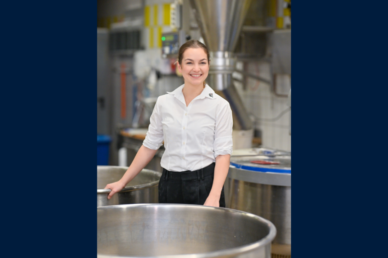
Foto van Berta Cillero Pastor

Here we are looking at the 'molecular map' of an infra-patellar fat pad (or Hoffa) patient's biopsy. An infra-patellar fat pad, also known as the Hoffa fat pad, is a specific type of adipose tissue located beneath the kneecap (patella) within the knee joint. It is an important component of knee anatomy, contributing to shock absorption and knee stabilization. The material is obtained thanks to a long-standing collaboration between the orthopaedic surgeons at the MUMC+, the orthopaedics laboratory, M4i and the MERLN institute.

Also read

  • On February 8, 2024, Ziemons received her PhD from Maastricht University for the dissertation “The role of the gut microbiota in human cancer, the power of an equilibrium". This dissertation is the result of Ziemons' research on the role of the microbiota in cancer – colorectal cancer but also...

  • Last November, Rory Koenen delivered his inaugural lecture following his appointment as a professor in 2023 at the research institute CARIM. The chair he holds is "Biochemistry of Vascular Inflammation and Thrombosis," and Rory's research can be best described as the quest to understand the role of...

  • judith sluimer

    Fresh air

    Vascular biologist Judith Sluimer was appointed professor of cardiovascular pathophysiology in October 2020. In her inaugural lecture, she alluded both to the importance of oxygen in the functioning of the heart and blood vessels as well as to the ‘fresh air’ she believes academia is always in need...