Brain atlas leads to fewer side effects in radiotherapy for brain and head and neck tumours
With the irradiation of tumours in the brain and in the head and neck area, the radiation dose can now be reduced considerably, without the desired treatment result being compromised. This drastically reduces the chances of side effects. Daniëlle Eekers, radiation oncologist at MAASTRO clinic and ZON-PTC was awarded her PhD on this subject last Friday, 7 December, at Maastricht University. She compared various treatment methods for her research. At her initiative, a brain atlas was developed at MAASTRO—together with the European Proton Centres and in close collaboration with Maastricht UMC+.
Remarkable
Before a tumour can be irradiated, scans (CT) are made to determine precisely where the correct dose should be given in relation to the location of the tumour. But it is often difficult to see on the scans where exactly the tumour is and where it is adjacent to the healthy tissue, for example with the optic nerve or the memory areas. These are also called organs at risk. That is why it is digitally contoured on very detailed scans (MRI) of the head of the patient. “In practice, there appeared to be important differences in how this was done by the various European Centres for Radiotherapy.” That is not really remarkable, according to Daniëlle Eekers, because this involves very complicated anatomy. She proposed the development of a brain atlas. Through a solid consensus, MAASTRO has now produced a definitive brain atlas that will be used by the European Proton Centres.
Brain atlas
The other European radiotherapy centres will also use this brain atlas. “It has become a digital atlas with images of the brain that is available online”, says Daniëlle Eekers. “You can view it from multiple perspectives, both on MRI and CT scans. The intention is that all radiotherapists will contour the many important organs at risk in the same way. This not only allows for good comparisons of the various treatments, such as the current irradiation techniques with radiation with charged particles (protons). But this also reduces the side effects of the radiation because it is now possible to take these organs at risk into account when determining the parameters of the radiation. The brain atlas thus makes an important contribution to the further optimisation of the radiotherapeutic treatment of tumours in the brain and the head and neck area.”
Cerebellum
Daniëlle Eekers also established that the cerebellum can play an important role in the recovery of the patient after radiation treatment. “It was already previously known that the cerebellum is primarily responsible for our coordination and balance. Now it appears that the posterior part of the cerebellum also affects the process of acquiring knowledge through perception and processing it through thinking. Sometimes patients have problems with this after radiation. It is therefore essential that the posterior part of the brain is carefully contoured to prevent unnecessary side effects.”
Protons
With two international studies, Danielle Eekers showed that the aforementioned organs at risk will receive lower doses by irradiating charged particles, such as protons and carbon. “The special thing about these particles is that they stop in the patient and therefore do not release a dose beyond the tumour.” This radiation with protons is now possible in Groningen and Delft and in a few weeks will also be in Maastricht. It is anticipated that approximately 3% of all patients who receive radiation therapy in the Netherlands are eligible for proton radiotherapy.
Source: press release MAASTRO clinic
Also read
-
Care for Climate, Care for Health: CAPHRI Research Day 2026
On June 10, 2026, CAPHRI held its annual Research Day. This year's urgent theme, climate health, highlighted the critical link between planetary and human well-being.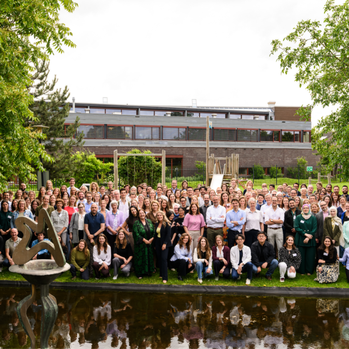
-
Pre-registration open for Education Research Day 2026
EDLAB warmly invites you to join us for a full day of sharing, learning, skill development and networking inspired by your passion for education research and innovation.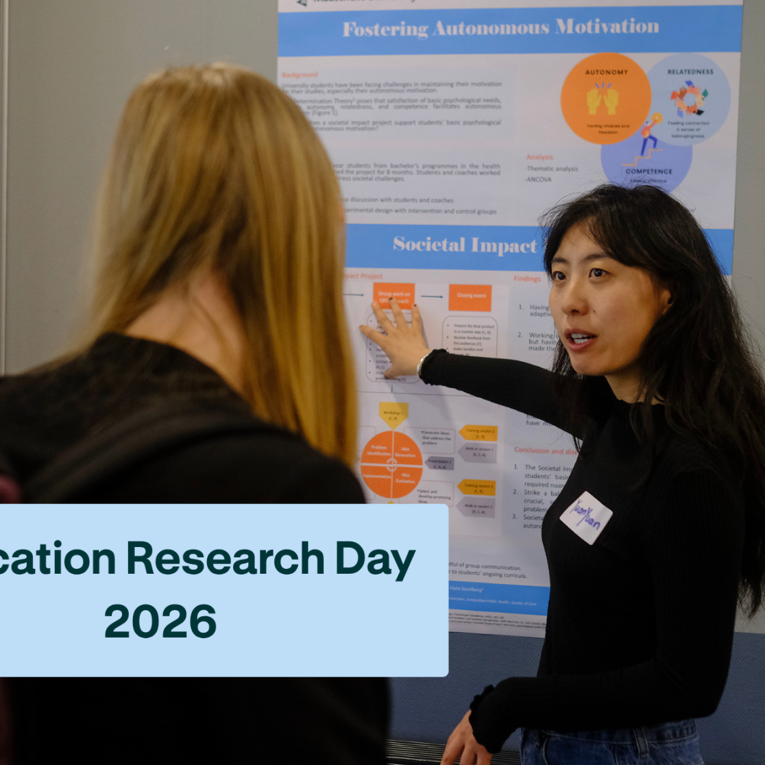
-
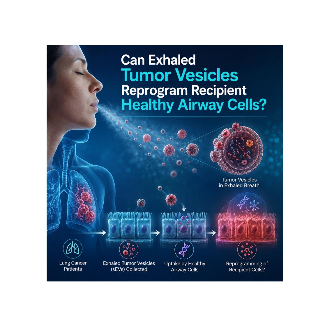
NWO-XS grant awarded for research on exhaled tumor vesicles
Congratulations: Double NWO-XS success on EVs-research NWO-XS grant awarded for research on exhaled tumor vesicles