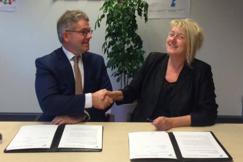
Dr. Benedikt Poser and his team reel in the NWO Investment Subsidy Medium grant (MaGW)
Last week Dr. Benedikt Poser of the Department of Cognitive Neuroscience (CN) at the Faculty of Psychology and Neuroscience (FPN) received word from the NWO (Dutch Organisation for Scientific Research) that his team received the NWO Investment Subsidy Medium grant (MaGW) from the NWO’s division of Social Sciences and Humanities. The grant of €453.200, with €151.100 additional co-funding by Scannexus BV, will be used to bankroll the investment project: “Improving reliability of high-field MRI in application to neuroscience and patients”
The aim of the FPN run project is to expand the capabilities of the 7 Tesla MRI scanner that is housed at the Scannexus MRI facilities at Oxfordlaan 55 in Maastricht. Benedikt Poser has been working on the development of brain imaging techniques for ultra-high field MRI for almost 12 years. He joined FPN four years ago where he is now the head of the MRI R&D group of Maastricht Brain Imaging Centre (M-BIC). His work continues to focus on the development of new pulse sequences and image reconstructions for ultra-high field MRI, and Maastricht’s 7T and 9.4T human MRI scanners.
“These high-field machines provide great opportunities for brain imaging, but there are also a bunch of challenges.” The challenges Poser and his team want to tackle with this investment project are head motion and the magnetic field fluctuations created when a person is inside the scanner. “Think of a big person lying in the scanner: when they breathe, their chest goes up and down and a lot of blood moves around which changes the magnetic field. This registers as intensity fluctuations on the received brain images and this will give a distorted view. When you’re trying to map brain activation with ‘functional MRI’, which is one of our primary applications, you are interested in measuring small local changes in blood flow signals over time. This gets really difficult if there is motion and the background signal is constantly changing. So we want the baseline to be as stable as possible, making it easier to pick up changes in brain activation. Field distortion and motion can also ruin anatomical scans, as these can easily take 5-10 minutes to acquire, and any breathing and motion will create a blurred image. It’s especially difficult with elderly subjects who are uncomfortable, patients who may be in pain, or kids, try telling them to lie still!”
The MaGW grant will be used to purchase a Field Camera System and an Open 32 Channel Head Coil.
Field Camera System
The field camera will make it possible to register those field fluctuations and use the data to “compensate” the image in post-processing as the simplest application. More advanced will be to record these fluctuations and feed the information into the scanners in real-time, to already make adjustments during the image acquisition. “I think this will improve almost every scan we make with the 7T, if we get it running as smoothly as I’ve boldly promised in the grant application!”
Open 32 Channel Head Coil to enable optical motion tracking
The head coil is the part of that scanner that fits around the subject and receives the signal. Normally these coils are closed, with only a small slit for the subject to see through and look at the screen. “We already have an optical camera system that can be mounted inside the scanner and allows you to track head motion. We place a little marker somewhere on the head and the camera detects it, and from that we can calculate the head motion. It is a beautiful system if it works, but realistically we’ve not really been able to use it because the coil has a closed design and the camera cannot see the marker. We plan to buy a specially designed head coil that has an open design, making it possible for the camera to see the marker. Moreover, the coil will be designed to incorporate the sensors of the field camera system. Together the field camera system on the one hand, and the open head coil for the optical motion tracking on the other, make a great comprehensive package. It gives us the armour we need to shield us from all unwanted nuisance effects that you encounter when you do MRI on people. The ultimate goal is to get the sharpest image possible from our scanner.”
Application
“In principle, all applications of MRI should benefit to some degree, even in our fMRI applications for cognitive neuroscience research where we often have the luxury of choosing experienced subjects who can lie still, or if necessary we just require the data. But think of patients. For example, I’m increasingly working with Dr. Heidi Jacobs of the Alzheimer Centrum Limburg, and here we do research on people with early onset Alzheimer’s or more advanced dementia. That is much tougher, these subjects or patients will move around at some point, and more often than not you can end up with data that is not useable. All patient populations are very precious, and we want to minimise the burden on them while getting the best possible data –so if we can increase the likelihood of a successful scan, even by a few percent, we will have won a lot!”
User-friendly
“The Middelgroot (MaGW) project is clearly a methodologically and technically very challenging project. But the express goal is to deliver implementations that will benefit the majority of our scanner users. But these people are “users”, and they should be able to just use the scanner for their research application, they don’t want to fiddle around with the extra gadgetry connect to the scanner. So far, most work with field cameras has been hardcore methods research, not so much targeted at real applications yet. I love methods research; it’s needed to keep pushing the boundaries of what is technologically possible. But when working with such technologies and the amazing MRI scanners we have here, I also always ask the question: how can we do it that people benefit from all this in real life, today and not just in five years. That is also why I work at FPN, where I’m mostly surrounded by users and not by other developers. The challenge now is to implement the set-up in a way that is user-friendly enough for people to say: I’m going to use this.”
Future directions?
Poser is also anxiously awaiting news on an NWO Vidi grant proposal that would maximally benefit from the field camera system. “It is not a coincidence that I submitted these proposal that the same time. For the Middelgroot, I want to improve standard imaging techniques that we already use here every day. The Vidi would allow me to do more fundamental methods research, and one aspect is on new and more efficient ways to acquire data using so-called non-Cartesian techniques. This involves driving the MRI scanners, specifically their gradients, to their absolute maximum so that one inevitably encounters system imperfections. Then you don’t know whether the system is really exactly doing what you ask of it. This is a problem if you want to reconstruct an image, it will be blurred. Tedious calibrations are one option, but if you have a field the camera then you’ll get all the required information exactly and on the fly, and you can feed that into the reconstruction to make a sharp image. So the Vidi proposal would benefit greatly from the field camera and really open up new opportunities; but well, we’ll just have to wait and see, and keep finger crossed for another few weeks!”
With special thanks to Dr. Benedikt Poser!
Also read
-
Centre of Expertise for Inclusive Labour Organisation (Expertisecentrum Inclusieve Arbeidsorganisatie)
On 7 December 2016 the UWV and Expertisecentrum Inclusieve Arbeidsorganisatie (CIAO) signed a cooperation agreement for the duration of 4 years. The CIAO is housed at Universiteitssingel 40, Maastricht University.

-
Dr. Jessica Alleva awarded for her work on improving body image
In January Dr. Jessica Alleva, from the Department of Clinical Psychological Science at the Faculty of Psychology and Neuroscience, was honoured twice for her work on improving body image. She received the 2016 Seymour Fisher Outstanding Body Image Dissertation Award and the Distinguished Women...
