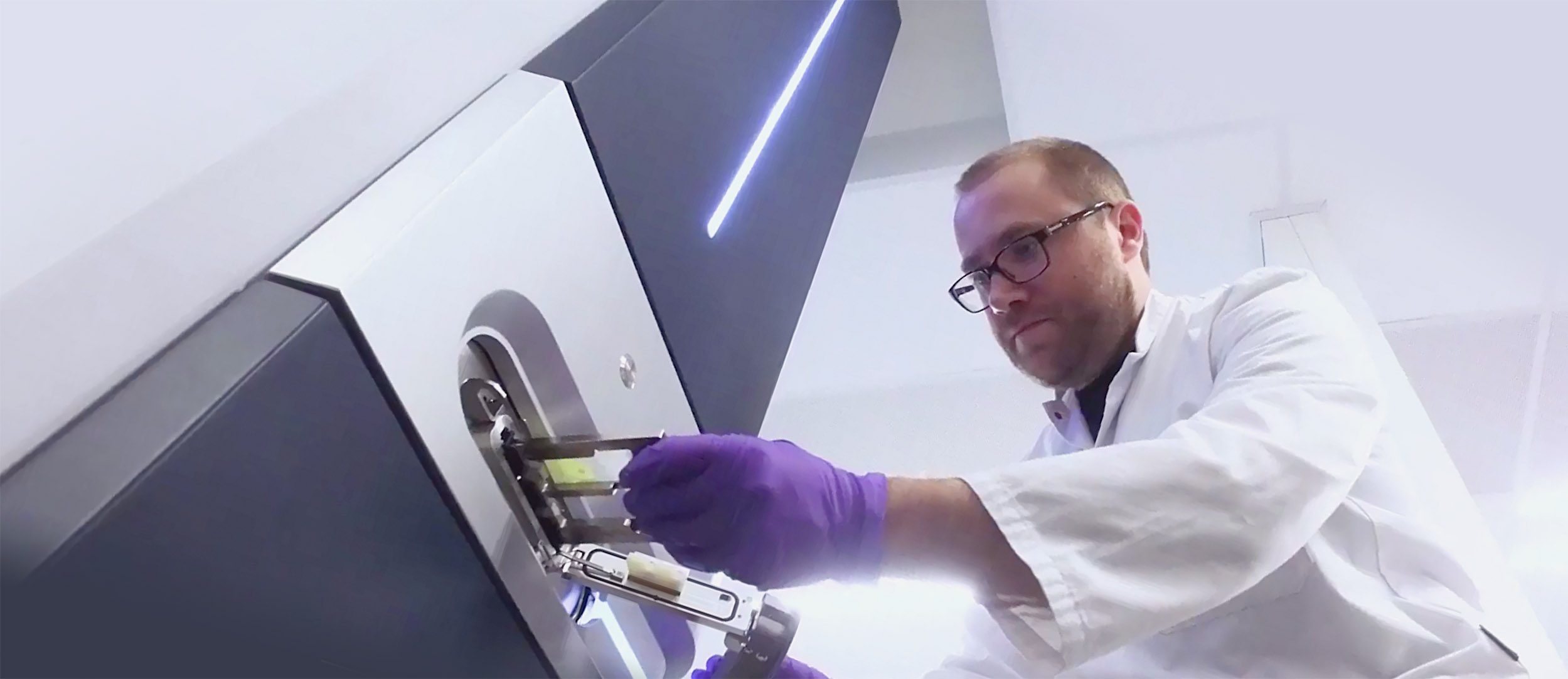
IMS infrastructure

M4I Division of Imaging Mass Spectrometry has the following capabilities:
The IMS CORE lab provides service, training, support and access to the MS imaging and proteomic technologies
Mass Spectrometry
- 9.4T MALDI/ESI SolariX Fourier Transform Ion Cyclotron Resonance imaging mass spectrometer
- 7T LTQ-FTICR system for ambient ionisation and imaging
- LAESI DP-1000 ambient imaging system of Protea Biosciences
- Bruker Ultraflex III ToF/ToF MALDI molecular imager with an IonPix Camera
- A Bruker RapiFlex TissueTyper
- A MS/MS enabled Bruker Rapiflex TissueTyper
- A Bruker MALDI-ToF Biotyper for microbial screening
- Physical Electronics NanoTof V Tandem MS SIMS system for high resolution SIMS equipped with a Bismuth, C60 and Argon Cluster sources
- A Physical Electronics TRIFT II for SIMS imaging with a Gold source
- TRIFT II based mass microscope for direct ion imaging with MALDI and C60-SIMS
- Waters 8K MALDI/ESI Synapt G2Si system for ion mobility enabled high resolution molecular imaging
- Waters 32K MALDI/ESI Synapt G2Si system for ion mobility enabled high resolution molecular imaging and MRM
- Waters MALDI Synapt HDMS system for conformational molecular imaging
- A Waters Xevo q-ToF system equipped with a DESI source for ambient ionization based imaging
- A Waters Xevo-q-ToF system equipped with a REIMS source for the development of intraoperative diagnostics
- A Waters HEPA filtered Xevo q-ToF systems system equipped with a REIMS source for research on intraoperative diagnostics
- A Waters TQS-micro triple quad with DESI-MRM capabilities.
- A Thermo Scientific q-Exactive HF system with a nano-LC system
- A Thermo Scientific Orbitrap Elite with a Spectroglyph MALDI imaging source for high resolution imaging MS
- A SimulToF MALDI Linear ToF imaging system
- An LCT q-ToF system modified for native MS and fitted with a high molecular weight IonPix Camera
Sample preparation
A fully equipped sample preparation facility, including cryosectioning, staining, analytical microscopes, matrix deposition and coating devices such as:
- Advion Triversa Nanomate system
- Several matrix deposition systems. ImagePrep, SunCollect and HTX sprayer
- 2 Cryo-microtomes for imaging MS
- Mirax histology scanner
- Leica optical research microscope with UV fluorescence imaging module
- ChIP spotting robot for LC-MALDI
- Virtual laboratory based image processing and analysis tools, including tissue classification and correlation software and protein data basing tools
- Large scale data storage and cluster computing facilities
Access to our facilities
Requests to access our Imaging Mass Spectrometry facilities at M4I can be specified using the 'Access to facilities'-contact form. The lab manager of the requested facility will evaluate your request.
M4I office wing
The M4I office wing has been designed with the same open and transparent look and feel as our labs. Based on C.O.R.E. collaborative open research education. C.O.R.E. requires a transparent and open environment for both laboratories and offices. M4I has invested heavily in an innovative and open environment for collaborative research. Research and office space is shared by scientist from very different backgrounds and disciplines, ranging from the fundamental sciences, technology and engineering as well as clinicians. In line with the CORE philosophy of Maastricht University the infrastructure is primed for researchers to cross the boundaries of their own disciplines and stimulate each other to excel in translational imaging science.
